
CT Checklist Before Using Any Hemorrhage Score
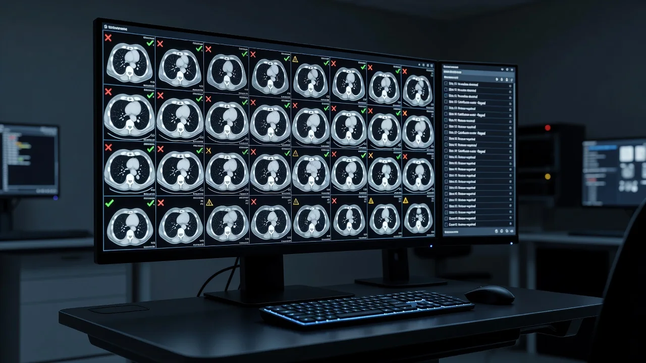
Accurate prognostication after spontaneous intracerebral hemorrhage depends on reliable imaging interpretation before any formal score is applied. Even well-validated tools such as the ICH score or FUNC score can mislead if the underlying CT findings are misinterpreted or incomplete. A structured visual checklist performed at the scanner console or PACS workstation improves consistency and prevents premature prognostic statements that may influence early care decisions.
Clinicians should systematically evaluate five key elements on the non-contrast CT: precise hemorrhage location, presence and extent of intraventricular hemorrhage, early signs of hydrocephalus, degree of mass effect, and technical adequacy of measurements. These features directly affect both mortality risk and functional outcome estimates. The treating team retains ultimate responsibility for integrating imaging data with clinical context and patient values.
Hemorrhage Location and Associated Features
Deep hemorrhages centered on the basal ganglia or thalamus typically arise from hypertensive arteriolosclerosis, while lobar locations raise suspicion for cerebral amyloid angiopathy, especially in patients over 55 years. Pontine or cerebellar bleeds carry distinct outcome implications because of brainstem compression risk. Documenting the epicenter and any extension into adjacent compartments prevents misclassification that could alter perceived prognosis. Location also guides decisions about neurosurgical consultation and blood-pressure targets.
Intraventricular Extension, Hydrocephalus, and Mass Effect
Intraventricular hemorrhage markedly worsens prognosis and is a core variable in most scoring systems. Even small amounts of blood layering in the occipital horns can obstruct cerebrospinal fluid flow. Look for ventricular enlargement out of proportion to age-related atrophy, effacement of cortical sulci, and midline shift exceeding 3 mm. Transtentorial herniation signs such as compression of the ambient cisterns should be reported immediately. These radiographic findings often drive urgent external ventricular drain placement before any formal score is calculated.
Accurate volume measurement is essential yet frequently performed incorrectly. The ABC/2 method remains practical at the bedside, but irregular shapes or multi-compartment bleeds require careful adjustment or planimetric software. Slice thickness, windowing, and motion artifact can each introduce error exceeding 10 mL, enough to change risk category in many validated models.
Practical CT Checklist
- Confirm hemorrhage epicenter (lobar, deep, brainstem, cerebellar)
- Quantify intraventricular blood (present/absent, Graeb or IVH score)
- Assess for hydrocephalus (temporal horn width, Evans index, sulcal effacement)
- Measure mass effect (midline shift in mm, cistern compression)
- Verify measurement quality (proper ABC/2 technique, adequate CT windows, no significant artifact)
| Feature | Imaging Sign | Prognostic Impact |
|---|---|---|
| Location | Lobar vs deep vs infratentorial | Alters etiology and surgical candidacy |
| Intraventricular hemorrhage | Blood in ≥1 ventricle | Increases mortality odds ratio >2 |
| Hydrocephalus | Enlarged ventricles + sulcal effacement | Predicts deterioration within 24 h |
| Mass effect | Midline shift >5 mm | Strong predictor of poor functional outcome |
| Volume accuracy | ABC/2 or planimetry | Each 10 mL increase raises mortality risk |
Using this checklist before applying any numeric hemorrhage score promotes intellectual honesty and reduces cognitive bias. It also creates a shared mental model for multidisciplinary discussions involving neurology, neurosurgery, and critical care teams. While no checklist replaces clinical judgment, systematic review of these CT elements helps ensure that subsequent prognostic conversations with families rest on firm radiographic ground. The final interpretation and management decisions always belong to the treating team.