
CT Measurement for ABC2-SPH Mortality Score
Calculating the ABC/2 method on a non-contrast head CT remains the fastest way to estimate intracerebral hemorrhage volume at the bedside. The result feeds directly into the ABC2-SPH mortality score, which combines hematoma volume, intraventricular extension, age, and admission Glasgow Coma Scale to give a realistic 30-day survival estimate. Getting the volume right matters: a 10 mL error can swing the predicted mortality by more than 15 percent.
Start by scrolling through the CT stack in brain window settings (width 80 HU, level 40 HU). Identify the slice that shows the largest area of the hematoma. This is your “C” slice. Measure the longest diameter (A) on that slice in centimeters, then the longest perpendicular diameter (B) on the same slice. Count the number of slices showing hemorrhage, multiply by slice thickness in millimeters, and divide by 10 to obtain “C”. The formula is simply (A × B × C) / 2. For irregular bleeds, many clinicians now use the “ABC/2 plus” modification that adds a second measurement on the slice showing the second-largest area, but the classic single-slice approach still performs well for the ABC2-SPH calculator.
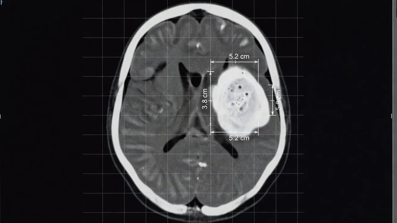
Choosing the Right Slice and Avoiding Common Pitfalls
Slice selection is where most errors occur. Never pick the topmost or bottommost slice if the bleed is tapering; these partial volumes underestimate the true diameter. If the hematoma spans more than five slices, double-check that you are counting every slice that contains any hyperdensity, including those with only a thin rim. Beam-hardening artifact near the skull base or calcified pineal can mimic blood; scroll up and down to confirm continuity with the main clot. In patients with multiple hemorrhages, measure only the largest one for the ABC2-SPH score unless the radiologist explicitly states otherwise.
Another frequent mistake is mixing units. All linear measurements must be in centimeters; slice thickness is entered in millimeters before dividing by 10. Rounding too early (for example, calling a 4.7 cm measurement “5”) adds unnecessary error. Digital calipers on modern PACS systems are accurate to 0.1 cm, so use that precision and round only the final volume to the nearest whole number.
Practical CT Checklist Before Calculating ABC2-SPH
Run through this quick mental checklist every time to keep your measurements reproducible:
- Confirm brain window settings (W80/L40) and no motion artifact on the chosen slices
- Identify the single largest axial cross-section for A and B
- Measure A and B perpendicular to each other, avoiding the longest oblique line
- Count every slice with measurable blood for C (include slice thickness correctly)
- Exclude extra-axial blood or chronic hypodensities from the volume
- Document intraventricular hemorrhage presence for the SPH component
- Double-measure if volume exceeds 30 mL or looks irregular
Turning Volume into Mortality Risk
Once you have a reliable ABC/2 volume, plug the numbers into the validated ABC2-SPH nomogram or online calculator. A 45 mL putaminal bleed with intraventricular extension in a 68-year-old patient with GCS 9 yields roughly 65 % predicted 30-day mortality. Recording both the raw volume and the final score in the radiology report helps the stroke team make faster, evidence-based decisions about blood-pressure targets, reversal agents, and family discussions.
| Volume (mL) | SPH Points | Approx. 30-day Mortality |
|---|---|---|
| <10 | 0 | 10-15 % |
| 10-29 | 1 | 25-35 % |
| 30-49 | 2 | 50-60 % |
| ≥50 | 3+ | >75 % |
Mastering the ABC/2 technique takes only a few supervised cases. After that, the entire measurement and checklist can be completed in under 90 seconds, giving clinicians a trustworthy number they can act on before the patient even leaves the scanner room.