
How ABC/2 Hematoma Volume Is Measured on CT
The ABC/2 method provides a rapid, practical estimate of intracerebral hematoma volume on non-contrast CT. It is widely used in emergency settings because it requires only a ruler and basic arithmetic, allowing clinicians to gauge severity, predict outcome, and guide decisions about blood-pressure control, reversal of anticoagulation, or surgical consultation. While not as precise as software-based volumetry, ABC/2 correlates well with formal measurements in most spherical or ellipsoidal hemorrhages.
The technique assumes the hematoma approximates an ellipsoid. Three perpendicular diameters are measured in centimeters, multiplied together, and divided by two. The result is expressed in milliliters (cm³). This quick calculation helps stratify patients for clinical trials and bedside prognostication, though final management decisions always belong to the treating team after integrating clinical context and serial imaging.
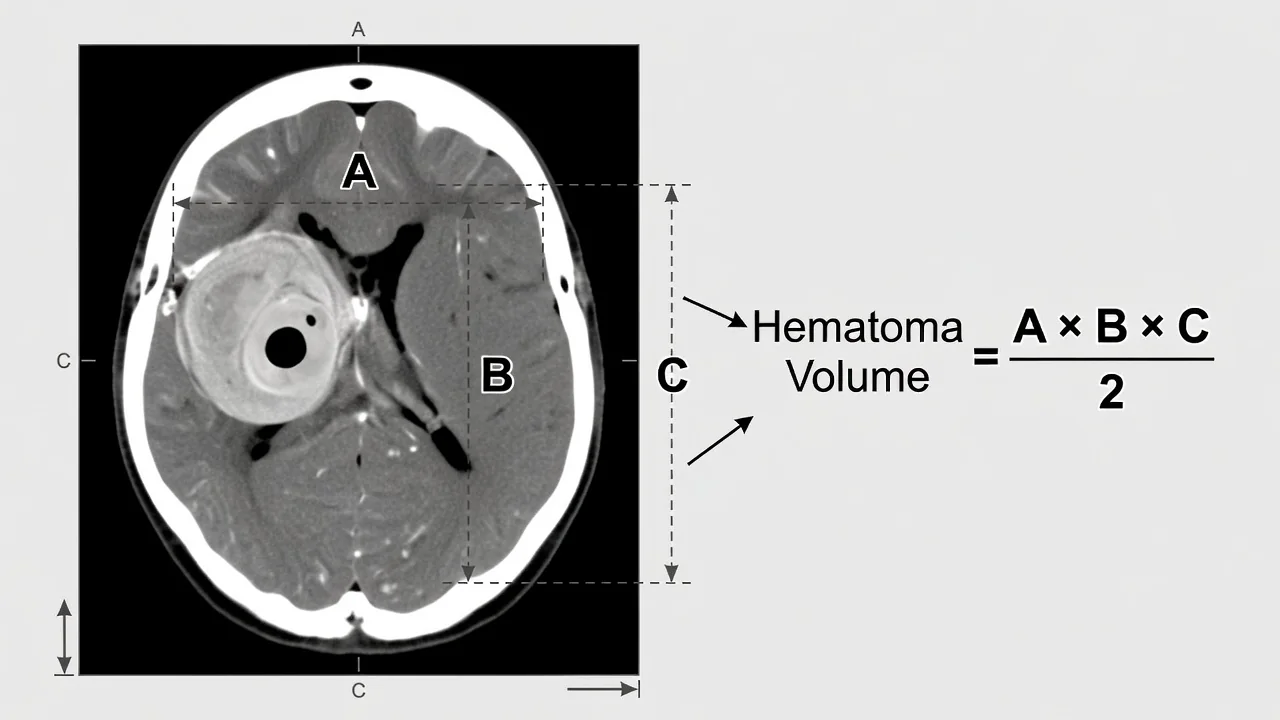
Measuring the Diameters
A is the largest diameter of the hematoma on the axial slice that shows the greatest area. Measure from the outer margin to the outer margin, ignoring thin surrounding edema. B is the largest diameter perpendicular to A on the same slice. Both A and B are obtained from the single slice judged to contain the largest cross-sectional area of the bleed.
C represents the vertical extent of the hematoma. Count the number of CT slices on which the hemorrhage is visible, then multiply by the slice thickness in millimeters and convert to centimeters. For example, if the bleed appears on 5 contiguous 5-mm slices, C equals 2.5 cm. When slices are not contiguous or the hematoma is irregularly shaped, experienced readers adjust by estimating the percentage of each slice occupied by blood before summing.
Common Pitfalls in Slice Selection
Selecting the wrong reference slice is the most frequent source of error. Clinicians sometimes choose the slice with the largest visible diameter rather than the slice with the largest cross-sectional area; these are not always identical. Another mistake is measuring C from the first to the last slice that touches even a sliver of blood, which overestimates volume when the hematoma tapers sharply at the poles.
Irregular, multilobulated, or intraventricular hemorrhages also challenge the ellipsoid assumption. In these cases, ABC/2 tends to overestimate true volume. When the hematoma spans markedly different slice thicknesses within the same study, or when metal artifact or beam-hardening obscures borders, the method loses reliability. In such situations the treating team should consider formal volumetric analysis using planimetric software.
When to Prefer Formal Volumetry
ABC/2 performs best for compact, supratentorial bleeds without substantial intraventricular extension. For research protocols, very large hemorrhages (>50 mL), or when precise volume is required to determine eligibility for minimally invasive evacuation, semiautomated or fully automated segmentation is preferred. Modern CT workstations and PACS tools can generate these measurements within seconds, reducing inter-observer variability.
Clinicians should still master ABC/2 because it remains the fastest bedside tool and is embedded in many guidelines and risk scores. Serial estimates using the same method allow rapid detection of hematoma expansion. When discrepancies arise between ABC/2 and clinical findings, or when surgical planning is contemplated, the treating team should obtain quantitative volumetry and consider repeat imaging.
Key points to document
- Pick the slice with the largest hematoma diameter for A.
- Measure B perpendicular to A on the same slice.
- Count C as the number of slices with visible hematoma multiplied by slice thickness.
| Component | Definition | Common Error |
|---|---|---|
| A | Largest axial diameter on slice with greatest area | Using longest visible diameter instead of greatest area |
| B | Largest perpendicular diameter to A on same slice | Measuring on a different slice |
| C | Number of slices with hemorrhage × slice thickness (cm) | Including slices with only trace blood |
| Volume | (A × B × C) / 2 | Forgetting to divide by 2 |
Accurate application of ABC/2 improves communication between emergency physicians, neurologists, neurosurgeons, and intensivists. It also helps families understand the scale of injury without requiring advanced software. With practice, most clinicians can estimate volume within 10–15 seconds, making the method an enduring cornerstone of acute stroke imaging interpretation.